Post by Admin on Dec 14, 2015 6:51:31 GMT -7
Radiographic findings of the spinous processes clinically back healthy warmblood horses
Initiation
Horse owners and riders put their horses increasingly suspected of having a spinal disease the veterinary examination before. One reason may be that spinal diseases win also in human medicine in importance and are widely used. In contrast to veterinary significance is increasingly given in human medicine and psychosomatic back pain. In any case, the awareness has increased for spinal diseases in the population in recent years and thereby a back problem is more common also in horses suspected.
Symptoms in horses that are associated with back pain, are usually rideability or derating. This is for the rider or owner often a big problem. Many want to cause her horse unnecessary pain and therefore know whether back pain are a possible cause for insubordination and performance degradation in question. In addition, spinal diseases may be associated with financial losses especially in sport horses. Today in equestrian sport and keeping a lot of money is invested, professional riders and breeders earn their livelihood and are thus dependent on the health of their horses. Even recreational riders want relaxed and unburdened their hobby. Not least at purchase examinations of horses back to the study comes to a growing importance. For veterinarians, it is therefore important to carry out a sovereign spinal examination and be able to interpret the findings obtained.
Thanks to great technical progress has for several years is an increasingly differentiated survey findings in horses with suspected spinal problems possible. By clinical examination (inspection and palpation including provocation sample) in conjunction with radiology, scintigraphy and ultrasonography a detailed diagnostic assessment can be carried out at the back diseased horse. However, it can also horses that show clinical signs of spinal disease, radiographic changes have. Here frequently raises the question of what clinical significance of the standard have different radiographic findings.
The X-ray examination of the spine is often desired especially in horses with higher value in the purchase examination. This radiological findings must be properly collected and interpreted in order to educate the owner or purchaser of the forecast for the later purpose of use of a horse can. In this context, the spinal investigation in connection with purchase examinations of horses become very important and often leads to legal disputes between buyers and sellers and / or kaufuntersuchendem veterinarian.
In order to give veterinarians for the evaluation of various radiographs help at hand, has an X-Commission consisting of experienced horse veterinarians, a guide developed in which a review of X-ray findings is given and can be obtained from the Society of Equine Medicine in Germany , In the X-ray guidance, some findings are listed on the spinous processes among many findings on the limbs.
Since only few X-ray studies with larger numbers were clinically back healthy horses, radiographs should be evaluated for this work within the framework created by purchase examinations. The aim was to examine the occurrence of radiological changes at the spinous processes in horses, which revealed no evidence of a painful spinal disorder in adspection, palpation and provocation samples. In addition, an assessment of their clinical importance should be made known to the frequency of individual radiographic findings.
Literature Overview
Spinal diseases are often diagnosed in horses. Although "back problems" are considered in horses both by riders and by veterinarians as an oppressing problem whose treatment is not satisfactory in many cases. The main reasons are that the exact diagnosis challenging and the clinical significance raised examination findings is difficult to interpret. A prerequisite to understanding back problems, is a good knowledge of the anatomy of the back.
The basics of the following compilation of the anatomy of the thoracic and lumbar spine and their joints, ligaments and muscles are the descriptions of Kadau (1991), nickel et al. (1992) and Wissdorf et al. (2004). If the details of these authors from those of other authors from, they are covered in detail with references. Works of other authors about specific anatomical issues are cited in the text.
Anatomy of the thoracic and lumbar spine
The construction of the horse spine reflects the special adaptation to the rapid locomotion on land. Stability outweighs free mobility and the spine forms the bony body axis.
The functions of the spine:
- Protection for the spinal cord and its nerve branches leaving
- Carrying the load of the trunk (especially the intestines), the neck
to support and head
- Recognition and Measurement origin for inserierende soft tissue (muscles and ligaments)
- When moving the pulse of the hind limbs on the other
To transfer body and to support other movements of the body (Remiger 1953 and HAUSSLER, 1999).
The spinal column consists of many individual bones (vertebrae, vertebrae).
All vertebra have a common basic shape, which is modified depending on the function in the various regions of the body.
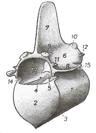
Fig. 1: Scheme half of a vertebra [Nickel et al. (1992)]
1. corpus vertebrae
2. Extremitas cranial
3. Crista ventralis
4. tape strip
5. veins hole
6. vertebral arch
7. spinous process
8. transverse process
9. articular process cranial
10. caudal articular process
11. processus mamillaires
12. processus accessoires
13. foramen vertebrae
14. vertebral notch cranialis
15. vertebral notch caudalis
The spine has seen from the side three curvatures:
- The dorsal convex head and neck curvature
- The dorsal concave neck Brustkümmung
- The dorsal slightly convex thoracic lumbar curvature
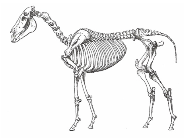
Fig. 2: The skeleton of a horse [Nickel et al. (1992)]
The thoracic spine
The thoracic spine of the horse is made up of 18 (17-19) together thoracic vertebrae. The vertebral bodies are short and average 5 cm long. The shortest is the 11th thoracic vertebra. From there they take cranial and caudal at length about something.
The thoracic vertebrae are characterized by the rib joint surfaces, which are formed by the costal foveae craniales et caudales. They are deep in the cranial region of the thoracic spine and caudal flat. At the last three vertebrae the fovea costalis cranialis merges with the fovea costalis transversi transversalis the processus.
The mobility of the vertebrae against each other decreases caudally toward. The reason for this is that the joint surfaces of the proc. articulares tangentially stand in the cranial region of the thoracic spine, more caudally they turn around and stand on the last two thoracic vertebrae sagittal (Jeffcott and DALIN 1980 TOWNSEND 1985 TOWNSEND and LEACH 1984. TOWNSEND et al 1983). From this area they are with the processus mamillary the processus mamilloarticulares merged.
The Extremitas cranialis and caudal are narrow and connected by epiphyses with the vertebrae.
The crista ventralis (Fig. 1) is 1-3. (4.) And 16 to 18. (15) thoracic vertebrae clearly formed. In the field of weak Crista ventralis of the 10th-15th Thoracic vertebra, i.e. in the saddle area, it can lead to overloading of new bone, osteophytes or exostosis z. B.. These proliferations can develop over two vertebrae as a bone bridge, which can lead to each other for complete fusion of the vertebral bodies.
The intermediate arc gap, space interarcuale, is the dorsal gap between two adjacent vertebral arches. By overlaying grasping the vertebral arches of the thoracic spine is missing here Spatia interarcualia.
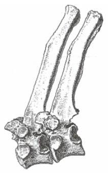
Fig. 3: 8 and 9 thoracic vertebrae of the horse [Nickel et al. (1992)]
Distinguish The thoracic vertebrae spinous processes are regions in shape, length, inclination angle and distance from each other. Kadau (1991) divides the thoracic vertebrae in type I (T3 to T9) and Type II (T13 to L6), where a Type I triangular cross-section, a pronounced Tuberosity, apophysäre cartilage caps and long spinous processes. Having inclination angle. Type II is more of a flat shape with längsovalem cross-section and wide spinous processes, resulting in a comb have thickened. In the central region prevail and wide distances between the ends of the Spinous close distances. The ends of the Spinous processes have a cranial Beak shaped tip and a broadened, sloping caudal area, the congruence for standing next vortex shows. The cranial edge of the spinous processes is narrow, while the caudal edge has a shallow groove or a crest and appear radiographically often doppellinig. In the middle part of the spinous processes, the periosteum is often rough, without the latter seems to have a clinical significance (Jeffcott, 1975b).
The cranial spinous processes of the thoracic spine form the withers. The first 5 spinous processes are increasing in length. The 6 spinous process is the highest (Jeffcott 1975b) and they will then gradually and rapidly shorter to 12 spinous process until 8 spinous process. Behind them are the same height with the spinous processes of the lumbar vertebrae. The spinous processes of slope in the cranial region to caudal and the caudal to cranial region. As anticlinal vertebra (the spinous process of this vortex is vertical) is usually the 15th, 14th or 16th of the rarely seen (NICKEL, 1947 and 1992; Jeffcott 1975b).
From the 3rd thoracic vertebra to the ends of the spinous processes to the spinous process are thickened tuberosity. The tuberosities bear in young animals cartilage caps of hyaline cartilage, have a private Verknöcherungskern and must are therefore considered apophyses. In withers the cartilage caps are widened 20-50 mm high and clearly, more caudally they are only a few millimeters high. The cartilage caps are formed with a half to one Year approached and have about 3 years achieved its final form. They show a significant Apophysenfuge which is closed only between 7 and 15 years old (Grimmelmann, 1977). The cartilage caps can grow in length, thus causing an increase in the withers.
The distances between the spinous processes were of Jeffcott and DALIN (1980) measured on carcasses. In the normal position of the spinous processes were in the range of T10 to T16 between 3.8 mm and 5.5 mm, in the region T16 to T18 an average of 8.2 mm, in the region T18 to L1 average 4.8 mm and in the range L1 to L2 average 11.0 mm measured. In the field of anticlinal vertebra passed the shortest distances: 3.4 mm between T14 and T15.
The lumbar spine
The horse has usually 6, less often 5 or 7 lumbar whose body bigger and longer than those of the thoracic vertebrae. The spinous processes are flat, the same length and tilt slightly to the cranial. Ventral to the vertebral body, there is a crest (Crista ventralis). The crista ventralis is no longer present on the last 2 to 3 vertebrae Extremitas cranialis and caudal are flat.
The transverse processes of the lumbar spine represent ribs rudiments and are termed processus costarii. The shortest lumbar transverse process is the first and the 3rd or 4th is always the longest. The edges of the transverse processes are sharp. Only at the last lumbar vertebrae, they are thickened towards the body and form the typical equine Intertransversalgelenke.
The cranial articular processes of the lumbar spine, as well as the last thoracic vertebrae, with the teats appendages to the processus mamilloarticulares merged.
The vertebral arches are high and so are the wide spinal canal. They are of the caudal articular processes, which spread to the base of the spinous processes, covered. This rarely Spatia interarcualia available. By interlinking the individual vertebrae into one another and the joint surfaces sagittal mobility of the lumbar spine is severely restricted. There is only one axial rotation possibility.
Joints of the spine
The joints of the spine are unique because each vertebra has two types of joints: both synovial and fibrokartilaginöse (HAUSSLER, 1999).
Joints between the vertebrae
Between the Extremitas cranialis and caudal vertebral no synovial joints but intervertebral joints (Symphyses intervertebrales) are formed. In this are the intervertebral discs and ligaments. longitudinal ventralis and dorsalis available.
The intervertebral discs are a bradytrophic tissue, ie, they are avascular and their metabolism is carried out exclusively by diffusion (BENNINGHOFF, 1985). Their outer zone of the annulus fibrosus consists of lamellar layered fibers, which are onion disk-like arranged and fused to the dorsal and ventral longitudinal ligament.
A special feature of the horse, the intervertebral discs are continuous fibrous and have at least macroscopically no nucleus pulposus (ROONEY 1979.1982; Jeffcott and DALIN, 1980; Kadau 1991 Dämmrich et al, 1993; TOWNSEND 1984.1986). In humans, the nucleus pulposus has a hydrous gelatinous core, its effect is considered to be shock-absorbing "incompressible water cushion" as very important. This importance of the horse must be considered highly unlikely because the nucleus pulposus is continuous fibrous (GRAY, 1973; LANZ and Wachsmuth 1982 BENNINGHOFF, 1985).
Small vertebral joints
The connections between the cranial and the caudal articular processes of the vertebrae that facet joints, represent synovial sliding joints, where the movement is parallel to the joint surfaces. Thus, the position of the facet joints affects the mobility of the spine. At the thoracic and lumbar spine, the joint capsules are progressively narrower inferiorly and the joint surfaces smaller. Accordingly, the mobility decreases caudally.
Townsend (1984) divided the thoracic and lumbar spine after mobility in four areas.
1. From T1 to T2
- Good dorsoventral flexion because large radial articular surfaces are possible available
- Also requires axial rotation and lateroflexion possible
2. From T2 to T17
- Pronounced axial rotation and lateroflexion possible, but only slight dorsoventral flexion because small tangentially directed joint surfaces exist.
3. From T17 to L6
- Zone of the lowest degree of mobility. All three types of movements are very restricted.
4. From L6 to the sacrum
- Place the greatest dorsoventral flexion possible by vertical joint surfaces, diverging spinous processes and thick pronounced intervertebral discs.
Spinal ligaments
Functionally can be attached to the backbone band two groups: The short spinal ligaments, which extend only between two consecutive vertebrae, and long spinal ligaments, which connect several vertebrae together.
Short spine bands
1. The ligaments. flava (slip sheet bands)
Close the Spatia interarcualia between the vertebral arches, made of elastic connective tissue and come as separate bands in the horse only between the 1st and 2nd thoracic vertebrae, the 5th and 6th lumbar and particularly strong in Spatium interarcuale lumbosacral ago (Kadau, 1991) ,
2. The ligaments. interspinalia (intermediate mandrel bands)
They are applied in pairs and fill in the spaces between the spinous processes of the thoracic and lumbar spine from. The intermediate mandrel belt is created in pairs and consists of five parts:
- The superficial
- The dorsal
- The center
- The ventral
- And a non-paired median portion connecting the sides each other contralateral

Fig. 4: Left view of an intermediate mandrel belt schematically [sketch accordingly HEYLINGS (1980)].
The fibers of the different parts of the intermediate mandrel bands combine scissor lattice-like to a plate. The majority of the fibers extends from the caudal edge of the front spinous obliquely kaudoventral the cranial edge of the following spinous process. The fibers of the dorsal part arise from connective tissue anterior to the ligament. Supraspinale, go to Interspinalraum and advertise at the cranial edge of the caudal spinous process.
An important role in the formation of ligaments. interspinalia play caudal to the 12th thoracic vertebra spinous process of the tendons of the spinal and caudally adjacent the longissimus dorsi, which the "functional" Lig. supraspinale form. They are attached not only to the cranial beak-shaped tips of the spinous processes and the same by the level of the falling to caudal summit surfaces, but also attract more deeply into the Interspinalräume into and form the dorsal parts of the ligaments. interspinalia.
3. The ligaments. intertransversaria (intermediate transverse process belts)
They extend between the transverse processes of two adjacent vertebrae and are really only at the origin of the transverse processes exist, since the space between the transverse processes is mainly filled by the strong sinewy interspersed quadratus lumborum.
Long spine bands
1. Lig. Nuchae (neckband)
consists of the neck and nape panel strand, both of which are formed in pairs.
The neck string, Funiculus nuchae, arises as a round strand at the external occipital protuberance, runs through the first cervical vertebra, connects above the 3rd cervical vertebra with the neck plate and then puts on the 4th, at partly on 3 spinous process of the thoracic spine. Here he joins the spinal ligament (Lig. Supra spinal).
The neck plate, lamina nuchae, springs with strong serrated dorsal crest of the Axis and the tubercle of the next three cervical vertebrae and the spinous process of the last two cervical vertebrae (Fig. 5). The cranial parts of the neck plate is connected to the neck strand and the side surfaces of the 3rd and 4th thoracic vertebrae spinous process, while the caudal parts only occasionally attach to the neck cord and principally for the spinous process of the 1st thoracic vertebra and the first Lig. Pull interspinous.

Fig. 5:.. History of a) and b nuchal ligament) Lig supraspinale the horse [Nickel et al. (1992)]
As a result of the influence of pressure is bursa between neck strand and Atlas (Bursa subligamentosa nuchalis cranialis) and between neck and strand Axiskamm (Bursa subligamentosa nuchalis caudalis) can form. Between the Widerristkappe and the 2nd and 3rd thoracic vertebrae spinous process is also often the Widerristschleimbeutel (Bursa subligamentosa supraspinalis).
2. The Lig. Supraspinale (back strap)
is the caudal continuation of the neck band and is from the 3rd-4th Thoracic vertebrae attached to the spinous processes of the following thoracic, lumbar and first sacral vertebrae.
According NICKEL et al. (1992), the band is back unpaarig and pulls a strand to the sacrum, the cranial part until the 15th-16th Thoracic vertebrae is elastic and then caudally is stringy. Also KOCH (1985) describes the back band as unpaired.
Kadau In 1991 the ligaments and joints of the thoracic and lumbar spine examined more closely and put on supraspinal ligament finds that the tape is to be divided into three sections:

Fig. 6: Classification of supraspinal ligament into 3 sections [from Kadau (1991)].
A. the elastic portion in the sloping area of the withers of 4/5.
until 9 / 10th thoracic vertebra
B. the paired elastic portion on the tendons of the muscles epaxial the 12th thoracic vertebra to just before the end of the thoracic spine (about 15.- 17th thoracic vertebrae). The spinous process of the 11th thoracic vertebra, the fascia of the spinal draws under the tape. For this reason, there is no firm anchorage between the tape and the spinous process peaks.
C. the purely sinewy portion (not "functional" as independent, but as Lig. supraspinal) starting on 12/13. Thoracic vertebrae, is formed by the tendons of the M. longissimus dorsi and M. spinalis and extends to the sacrum. This part is not continuation of the continuous elastic neck-back band, but overlaps with the under b. described structure on the posterior thoracic spine.
In the whole area of the thoracic spine, the back band is edged by the thoracolumbar fascia, which springs both lateral sides of the spinous processes.
The back band also bridges the space between L6 and S1 lumbosacral Spatium, and so affects the mobility of the Ileosakralgelenkes, especially the tilting of the pelvis.
3. The Lig. Longitudinal ventral (ventral longitudinal ligament)
is located on the ventral side of the vertebral body and is developed in the cranial thoracic vertebrae area only weakly. Next caudal to the last 8-9 thoracic vertebrae and the lumbar vertebrae, it is however strongly developed (MARTIN, 1914 SISSON and Grossman, 1975). It combines adjacent vertebrae together, and it comes in firm contact with the intervertebral discs.
4. The Lig. Longitudinal dorsal (dorsal longitudinal ligament)
runs at the bottom of the entire spinal canal from the dens of the 2nd cervical vertebra to the sacrum. It is attached to the rough block the dorsal surface of the vertebral bodies and the intervertebral disks, where it is connected to the outer fibers of the annulus fibrosis fixed (ELLENBERGER and BAUM, 1977).
The muscles of the spine
Jeffcott and DALIN (1980) divide the main muscles of the horse's back into three groups:
superficial muscles:
- Trapezius
- M. cutaneous
deeper muscles:
- Serratus dorsalis cranialis M.
- Serratus dorsalis caudalis M.
- M. longissimus dorsi
- M. multifidus dorsi
- Iliocostalis dorsalis
- M. intertransversalis lumborum
sublumbale and medium gluteal muscles:
- Psoas minor
- Psoas
- Iliacus
- Quadratus lumborum
- M. gluteals medialis
The major back muscle is the longissimus dorsi (Jeffcott and DALIN, 1980), consisting of a large number of relatively small segments and acts as the strongest extensors of the back and loin. The main task of this muscle is to ensure the stability of the spine during movement (Jeffcott and DALIN, 1980).

Biomechanics
The back is the central part of the musculoskeletal system of the horse and therefore extremely important for athletic performance. The diagnosis "back problems" is now placed more often than before. However, it is debatable whether this is due to an actual increase in the incidence of this disease or to the growing awareness of the problem "back disease" (VAN Weeren, 2004).
To understand the cause of back problems, as well as to investigate the circumstances that induce pain, be accurate knowledge, not only of the anatomy of the back, but also of the biomechanics of the thoracic and lumbar spine needs (DENOIX, 1999).
The term "Biomechanics" is understood to living structures, the application of the laws of mechanics. For area biomechanics heard the dynamics, which in turn is divided into kinematics and kinetics. The kinematics explains the movement of the body and the kinetics explains the change in the state of motion of bodies by forces acting (BADOUX, 1975).
Construction of the back / spinal functional structure
ZSCHOKKE (1892) compares the horse's back with a bridge and put the theory of vertebral bridge before, to explain the biomechanics of the horse's back. The limb should serve as a bridge pillars on which the forces are routed. The thoracic and lumbar spine with muscles and ligaments represents the bridge.
From Slijper (1946) the biomechanics of the horse spine was declared because of the similarity as a very flat Bowstring Bridge.

Fig. 8: Schematic representation of the principle of Bowstring Bridge to Slijper (1946).
When bowstrings bridge vertebrae and intervertebral discs conform to the flameproof "bow". Dorsal to the arch form spinous processes and vertebral arches the bony insertion of the ligaments. interspinalia, the Lig. supraspinal and the longissimus, the M. spinalis and the segmental Mm. multifidus. The "string" of the bridge arch form the abdominal muscles, particularly the rectus abdominis muscle. The tension of the sheet is effected by the intercostal muscles and the abdominal muscles. The dorsalkonvexe curvature arises primarily from the tension of the bowstring (= abdominal wall muscles). Lowering and voltage of the bridge arch (= spinal column), however, are the result of the contraction of the posterior muscles of the spine with the pulling direction according to kaudodorsal.
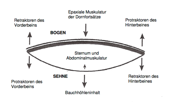
Fig. 9: Factors that influence the movement of the back according to the concept "arc and chord" [VAN Weeren (2004)].
The bowstring bridge is suspended superiorly by the Mm. serratus ventralis thoracis between the shoulder blades and rests inferiorly on both Capita femores so that the pelvic girdle is included. Thus, the thrust transmission of the hindquarters is guaranteed to the hull. The forelimbs are not directly of locomotion, but this support only by Stemmung and support the torso. The purpose of the indirect synsarkotischen compound of the forelimbs to the hull is in the shock absorption which is intended to prevent the vibrations while placing the limb are transferred to the brain (Koch, 1985).
While the function of the dorsal spinal muscles present numerous findings, is the function of the head and neck, the less considered on the neckband and the ligamentum supraspinal to the voltage conditions in the thoracic and lumbar spine partake (Jeffcott, 1979c;. ROONEY, 1979 1982).
In the literature, the head and neck are considered console that neutralized when pressing forward thrust forces occurring at the front end of the spine or displaced by raising and lowering the head of the center of gravity. It is obvious that the head and neck on neck band and Lig. Supraspinale are instrumental in the maintenance of the elastic stress of the spine. This applies to the dorsalkonvexe spinal curvature and even more for the extension of the spine. The latter is stabilized by contraction of the posterior spinal muscles with kaudodorsaler pulling direction, said kraniodorsal directed train of the lever head and neck supports the voltage. The voltage can be increased by extension of the lever arm. The head and neck accounts for about 30% of the total weight of horses. This is the significance of the head and neck for the tension evident (Dämmrich and RANDELHOFF 1993).
The tension of the bowstring design can be adapted to any posture and each movement phase (Jeffcott, 1979c). The mobility of the body axis is nuchal possible by the elasticity of the intervertebral discs, the Interspinalbänder and other intervertebral ligaments and the ligament.. The epaxial muscle plays a crucial role (SCHMALTZ 1928; Jeffcott, 1979c).
FAUQUEX confirmed with his investigations in 1982 that the changes in inventory between the spinous processes also by the attitude of the head and neck, as well as the position of the limbs are substantially affected. The viability of the horse's back can therefore bridge with theories, which include only the thoracic and lumbar spine can not be explained satisfactorily (FAUQUEX, 1982).
Acting on the spinal column
The spine of the horse is exposed to constantly forces. While standing, the spine has to bear the weight of the body, which means that the total mass of the horse on its center of gravity (CM) as a force is applied. The CM is a horse, which charged all four limbs equally, about 18 cm caudal to the xiphoid cartilage (KOCH, 1985). The power of body mass on gravity bends the spine anteriorly. To resist this bending, other forces must act against it (Rooney, 1979).
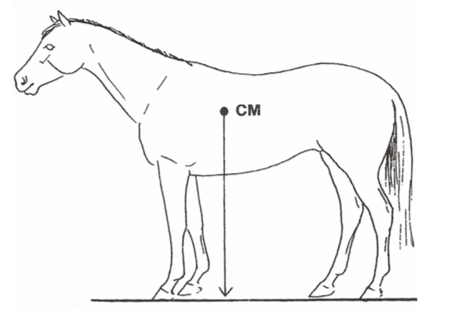
Fig. 10: "Center of Mass" (CM) = center of gravity of the body mass of the horse [ROONEY (1982)].
By the directed caudally train the back muscles - longissimus and Mm. multifidi - and directed cranially train the neck muscles - Mm. spinales dorsi (thoracis et cervicis) - the vertebrae against each other pressed. This leads to a stabilization of the spine and acts as the dorsoventral directed forces against (Rooney, 1979).
ROONEY (1979) presented this theory based on vectors represent in parallelograms of forces. For example, should the muscle power that counteracts the force of gravity, made up of two different components together, who work at a certain angle to the spine and can be analyzed by vectors.
The definitions of posterolateral and ventroflexion be given by various authors in the opposite fashion. Below based on Jeffcott and DALIN (1980) is designated dorsoflexion the lowering of the back and with the ventroflexion Aufkrümmung the back.
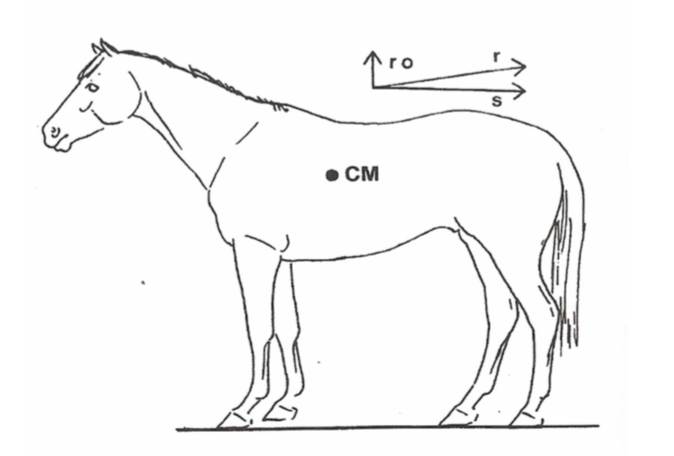
Fig. 11: Schematic representation of the costs associated with the caudal musculature of the horse's back force vectors [ROONEY (1982)].
The sacrum is seen as a fixed point and the back muscles exert characterized in contracting a train r after kaudodorsal (Fig. 11). This force represents a vector is and can be divided into two vectors: vector ro, pulling toward dorsal, and vector s, the caudal drifting towards. Here counteracts ro dorsoflexion the spine by lifting the spine posteriorly. Vector s represents the train, which presses the vertebrae against each other, what other way the spine is stabilized (ROONEY, 1979; Rooney, 1982).
Considered Spiegelverkehrt affects the back muscles with the forelimb as a fixed point as against the dorsoflexion spine. Both directed posteriorly forces r, on the one hand the strength of the dorsal musculature and on the other hand the strength of the neck muscles, act together against the pulling down of gravity and thus the dorsoflexion spine counter (ROONEY, 1979; Rooney, 1982).
If a horse is moving slowly, the back muscles, the tension is not maximally contracted, thereby not a maximum, which allows an increased flexibility of the spine. When the horse moves faster, increase the contraction forces in the back muscles, the vertebral bodies are solid pressed against each other and the spine is rigid and inflexible. (Jeffcott, 1979c, 1980; SLIPJER 1946 ROONEY, 1982) This is an effective starting point for the increasing driving force of the hind limbs developed.
The mobility of the thoracolumbar spine
The movement in the horse's back is the sum of each movement in the vertebral joints. Each vertebra has three joints: the intervertebral disc and on both sides of the facet joints. This design was created by Townsend and LEACH (1984) "Three-joint complex" called. Since the horse is the only animal species in which the Procc. transversi in the lumbar spine also articulate with each other, the authors are talking about a "Five-joint complex".
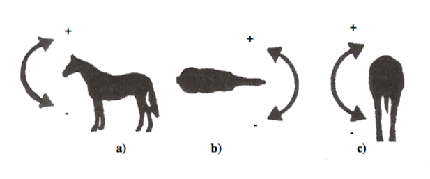
Fig. 12: AC: The basic movements of the back of horses:
a) + = diffraction (= ventroflexion) - = stretching (= dorsoflexion), b) lateral flexion and c) the axis of rotation [VAN Weeren (1982)]
The possibility of movement of each vertebra is explained here in the orthogonal coordinate system.
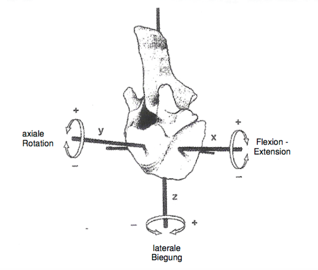
Fig. 13: Schematic representation of the basic movements of the spine represented by the rotation of a single vertebra within the 3 axes of an orthogonal coordinate system [VAN Weeren (2004)]
The ventroflexion and dorsoflexion spine are represented as rotation about the x axis. The rotation about the longitudinal axis of the spinal column is equal to the rotation around the y-axis. The lateral bending of the spine is represented by the rotation about the z-axis. Dorsoventral motion and axial rotation can take place independently of each other. The lateral deflection, however, can only be carried out in combination with the opposite axial rotation (Townsend, 1985).
The six movements run on the intervertebral from:
1. Longitudinal bend in the vertical plane of the flexion or extension of the spine is used (dorsoventral flexion or extension)
2. Transverse bending in the horizontal plane, which serves to turn the fuselage left or right (lateral flexure)
3. Rotation around the longitudinal, horizontal axis and rotation of adjacent vertebrae (axial rotation)
4. TransversaleScherung
5. LongitudinaleKompressionoderStreckungdesAchsenskeletts
6. VertikaleScherung
Based on these possible movements the intervertebral joints can be divided into four main groups.
T1-T2:
The first thoracic intervertebral joint is characterized by large dorsoventral movement but little axial rotation. The reason for this is that the caudal articular surfaces of T1 and T2 cranial articular surfaces of interlocking, which allows a dorsoventral movement, but no axial rotation. The spinous process of T1 is short. Since the nuchal ligament. Not inserted at T1, there is only a weak connection to the ligamentous T2. The intervertebral disc these two vertebrae is the strongest of the thoracic spine. This results in an increased dorsoventral mobility in this joint (Townsend, 1986).
T2-T16 :
Hier sind die Gelenkflächen schmaler, relativ flach und tangential in ihrer Ausrichtung. Sowohl dorsoventrale Bewegung, als auch laterale Biegung und axiale Rotation sind möglich (TOWNSEND und LEACH, 1984). Die dorsoventrale Bewegung in diesen Gelenken ist relativ eingeschränkt, was durch die dünnen ovalen Zwischenwirbelscheiben und die starken Verbindungen durch die Bänder zwischen den Dornfortsätzen (Ligg. interspinalia) bedingt ist (TOWNSEND, 1986). The lateral bending and axial rotation are significantly different in these joints. The joints between T9-T14 show this respect the greatest possible freedom of movement (Townsend and Leach, 1984). Jeffcott and DALIN Write (1980), that after a significant lateral bending T13 does not happen again and that caudal to T11 hardly takes place rotation.
T16-L6:
In the caudal thoracic and lumbar spine, the radial leads
Orientation of the intervertebral joints and the presence of articular processes, the. With the Procc mamillary are fused to the fact that only a small movement is present. TOWNSEND and LEACH (1984) studied the spine of 17 horses and found in 88% of animals Lateralgelenke between the transverse processes of L4 and L5. Of these 23% were ossified joints. Between L5-L6 and L6-S1 Lateralgelenke were formed in all animals, of which 59% from L5-L6 were fused. The Lateralgelenke limit the axial rotation and lateral bending.
L6-S1:
In this joint, the lumbosacral junction, find the greatest dorsoventral flexion and extension of the whole spine instead (SCHMALTZ 1928; SLIPJER 1946; Jeffcott, 1977; Jeffcott and DALIN, 1980 TOWNSEND et al, 1983;. TOWNSEND and Leach, 1984 ). The anatomical reasons are the small articular surfaces, the thick intervertebral discs, the great distance between the spinous processes of the vertebrae L6 and S1 and the little educated interspinous ligament tissue (Jeffcott and DALIN, 1980; Kadau, 1991). Rotation and lateral bending which are restricted by the engagement of the joint surfaces, the oval intervertebral discs and the presence of the Lateralgelenken (Townsend, 1986).
Tab. 1: Agility in different areas of the thoracic and lumbar spine of the horse (Townsend, 1985).

In a study by Johnson and Roethlisberger HOLM (2004) on the back kinematics in healthy horses has been found that the lumbar region at the Dressage is often longer than the jumping. Moreover Dressurpferde reported compared to jumpers on increased lateral mobility of the spine. Next mares showed a higher lateral mobility in the cranial thoracic region as geldings and older horses less flexion and extension of the back at a trot than younger horses.
Licka and PELHAM (1998) show in their study of horses standing flexibility in the back in clinically normal animals. The greatest mobility pointed T16 on. The flexibility of the back up and down has been expressed as a percentage to the withers. The average ventroflexion was T16 5.9%, the average dorsoflexion -2.4%. The average lateral flexion to the left was 4.2% and 5.3% to the right. Furthermore, the study showed that the lateroflexion the back always occurs in connection with a slight elongation of the spine.
Already from the earliest textbooks and representations of riding horses reveals that in the training of horse and rider in addition to the maneuverability of the horse and its capability has been sought to carry. This makes hooking the straps and Faszienplatten that, from occiput over the neck, the withers the back and the croup to reach down to the heel bone to the ankle of the horse, an essential role. The ability to support is by stretching and arching of the neck and spine and the lower contact of the hind quarters reached (FAUQUEX, 1982). The lowering of the head with a resulting voltage of the Lig. Nuchae always causes ventroflexion spine (DENOIX, 1980). The lifting of the head on the other hand leads to a dorsoflexion in the back. These relationships are of great importance in the formation of a dressage horse (VAN Weeren, 2004).
The raising and lowering of the back shows a double-sinusoidal movement pattern in the walk and trot and an easy-sinusoidal at a gallop (VAN Weeren, 2004). The movement of the spine is substantially smaller than in the walk and canter trot.
Back movements
In the posterolateral and ventroflexion the back the position of the vortex is changed. Angel Point, the small vertebral joints. As described above, the dorsally directed Aufkrümmung the back is done by contraction of the abdominal wall muscles (= tension of the bowstring). The spinous processes away from each other, but are limited by the Interspinalbänder in their distance. When Aufkrümmung the spine, the vertebrae closer to each other, the intervertebral disc (especially ventral) serves as druckaufnehmendes cushion and as a sliding layer. The sliding of the vertebral bodies to each other is also supported by the shape of the vertebral body extremities that are shaped like the head and pan.
In the ventral directed dorsoflexion the back the movement extension is not so great. Unlike Aufkrümmung the spinous processes approach in lowering the back at each other, the vertebral bodies are removed (especially ventral) and longitudinal ventral be by the Lig. Fixed. The intervertebral disc is especially dorsal pressure loaded (Dämmrich and RANDELHOFF, 1993).
The spine is similar to a "complicated assembled, elastic rod" which is maintained by passive structures in its shape. This bar - starting at the occiput - has an elastic connection down to the pulse-emitting hind legs. A prime example is the under the rider in complete suppleness going, gathered Dressage, in which the hind legs is strongly slid under the hull, so as to relieve the forehand maximum (FAUQUEX, 1982).
In its tendency can be the significance for riding movements of the horse's back and the horse's rump with six phases of motion describe (Hübener, 2004):
1. The withers are raised in the stance phase of / both forelimbs (the cannon bone is vertical).
2. The croup is raised in the stance phase of / both hindlimbs (the cannon bone is vertical).
3. The body of the horse swings in locomotion each side of the load-bearing, load supporting or load forward Temme ends hind leg.
Change 4. In step upward and downward movement of the withers and croup in opposite directions; This takes place in a sequence of movements twice (double- sinusoidal movement pattern).
5. decline in trot and rising withers and croup simultaneously. In the two phases of the suspension movement following the fifth position respectively reached their highest point (just-sinusoidal movement pattern).
6. In the gallop the withers at the stage of Einbeinstütze hindlimb and the croup achieved at the stage of Einbeinstütze the forelimb a climax. The saddle position achieved in limbo phase its highest point, the withers and the croup are at this moment on a plane.
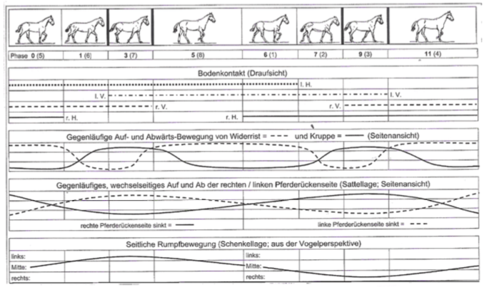
Fig. 14: footfall, ground contact and pelvic movements in the horse in the step
Initiation
Horse owners and riders put their horses increasingly suspected of having a spinal disease the veterinary examination before. One reason may be that spinal diseases win also in human medicine in importance and are widely used. In contrast to veterinary significance is increasingly given in human medicine and psychosomatic back pain. In any case, the awareness has increased for spinal diseases in the population in recent years and thereby a back problem is more common also in horses suspected.
Symptoms in horses that are associated with back pain, are usually rideability or derating. This is for the rider or owner often a big problem. Many want to cause her horse unnecessary pain and therefore know whether back pain are a possible cause for insubordination and performance degradation in question. In addition, spinal diseases may be associated with financial losses especially in sport horses. Today in equestrian sport and keeping a lot of money is invested, professional riders and breeders earn their livelihood and are thus dependent on the health of their horses. Even recreational riders want relaxed and unburdened their hobby. Not least at purchase examinations of horses back to the study comes to a growing importance. For veterinarians, it is therefore important to carry out a sovereign spinal examination and be able to interpret the findings obtained.
Thanks to great technical progress has for several years is an increasingly differentiated survey findings in horses with suspected spinal problems possible. By clinical examination (inspection and palpation including provocation sample) in conjunction with radiology, scintigraphy and ultrasonography a detailed diagnostic assessment can be carried out at the back diseased horse. However, it can also horses that show clinical signs of spinal disease, radiographic changes have. Here frequently raises the question of what clinical significance of the standard have different radiographic findings.
The X-ray examination of the spine is often desired especially in horses with higher value in the purchase examination. This radiological findings must be properly collected and interpreted in order to educate the owner or purchaser of the forecast for the later purpose of use of a horse can. In this context, the spinal investigation in connection with purchase examinations of horses become very important and often leads to legal disputes between buyers and sellers and / or kaufuntersuchendem veterinarian.
In order to give veterinarians for the evaluation of various radiographs help at hand, has an X-Commission consisting of experienced horse veterinarians, a guide developed in which a review of X-ray findings is given and can be obtained from the Society of Equine Medicine in Germany , In the X-ray guidance, some findings are listed on the spinous processes among many findings on the limbs.
Since only few X-ray studies with larger numbers were clinically back healthy horses, radiographs should be evaluated for this work within the framework created by purchase examinations. The aim was to examine the occurrence of radiological changes at the spinous processes in horses, which revealed no evidence of a painful spinal disorder in adspection, palpation and provocation samples. In addition, an assessment of their clinical importance should be made known to the frequency of individual radiographic findings.
Literature Overview
Spinal diseases are often diagnosed in horses. Although "back problems" are considered in horses both by riders and by veterinarians as an oppressing problem whose treatment is not satisfactory in many cases. The main reasons are that the exact diagnosis challenging and the clinical significance raised examination findings is difficult to interpret. A prerequisite to understanding back problems, is a good knowledge of the anatomy of the back.
The basics of the following compilation of the anatomy of the thoracic and lumbar spine and their joints, ligaments and muscles are the descriptions of Kadau (1991), nickel et al. (1992) and Wissdorf et al. (2004). If the details of these authors from those of other authors from, they are covered in detail with references. Works of other authors about specific anatomical issues are cited in the text.
Anatomy of the thoracic and lumbar spine
The construction of the horse spine reflects the special adaptation to the rapid locomotion on land. Stability outweighs free mobility and the spine forms the bony body axis.
The functions of the spine:
- Protection for the spinal cord and its nerve branches leaving
- Carrying the load of the trunk (especially the intestines), the neck
to support and head
- Recognition and Measurement origin for inserierende soft tissue (muscles and ligaments)
- When moving the pulse of the hind limbs on the other
To transfer body and to support other movements of the body (Remiger 1953 and HAUSSLER, 1999).
The spinal column consists of many individual bones (vertebrae, vertebrae).
All vertebra have a common basic shape, which is modified depending on the function in the various regions of the body.

Fig. 1: Scheme half of a vertebra [Nickel et al. (1992)]
1. corpus vertebrae
2. Extremitas cranial
3. Crista ventralis
4. tape strip
5. veins hole
6. vertebral arch
7. spinous process
8. transverse process
9. articular process cranial
10. caudal articular process
11. processus mamillaires
12. processus accessoires
13. foramen vertebrae
14. vertebral notch cranialis
15. vertebral notch caudalis
The spine has seen from the side three curvatures:
- The dorsal convex head and neck curvature
- The dorsal concave neck Brustkümmung
- The dorsal slightly convex thoracic lumbar curvature

Fig. 2: The skeleton of a horse [Nickel et al. (1992)]
The thoracic spine
The thoracic spine of the horse is made up of 18 (17-19) together thoracic vertebrae. The vertebral bodies are short and average 5 cm long. The shortest is the 11th thoracic vertebra. From there they take cranial and caudal at length about something.
The thoracic vertebrae are characterized by the rib joint surfaces, which are formed by the costal foveae craniales et caudales. They are deep in the cranial region of the thoracic spine and caudal flat. At the last three vertebrae the fovea costalis cranialis merges with the fovea costalis transversi transversalis the processus.
The mobility of the vertebrae against each other decreases caudally toward. The reason for this is that the joint surfaces of the proc. articulares tangentially stand in the cranial region of the thoracic spine, more caudally they turn around and stand on the last two thoracic vertebrae sagittal (Jeffcott and DALIN 1980 TOWNSEND 1985 TOWNSEND and LEACH 1984. TOWNSEND et al 1983). From this area they are with the processus mamillary the processus mamilloarticulares merged.
The Extremitas cranialis and caudal are narrow and connected by epiphyses with the vertebrae.
The crista ventralis (Fig. 1) is 1-3. (4.) And 16 to 18. (15) thoracic vertebrae clearly formed. In the field of weak Crista ventralis of the 10th-15th Thoracic vertebra, i.e. in the saddle area, it can lead to overloading of new bone, osteophytes or exostosis z. B.. These proliferations can develop over two vertebrae as a bone bridge, which can lead to each other for complete fusion of the vertebral bodies.
The intermediate arc gap, space interarcuale, is the dorsal gap between two adjacent vertebral arches. By overlaying grasping the vertebral arches of the thoracic spine is missing here Spatia interarcualia.

Fig. 3: 8 and 9 thoracic vertebrae of the horse [Nickel et al. (1992)]
Distinguish The thoracic vertebrae spinous processes are regions in shape, length, inclination angle and distance from each other. Kadau (1991) divides the thoracic vertebrae in type I (T3 to T9) and Type II (T13 to L6), where a Type I triangular cross-section, a pronounced Tuberosity, apophysäre cartilage caps and long spinous processes. Having inclination angle. Type II is more of a flat shape with längsovalem cross-section and wide spinous processes, resulting in a comb have thickened. In the central region prevail and wide distances between the ends of the Spinous close distances. The ends of the Spinous processes have a cranial Beak shaped tip and a broadened, sloping caudal area, the congruence for standing next vortex shows. The cranial edge of the spinous processes is narrow, while the caudal edge has a shallow groove or a crest and appear radiographically often doppellinig. In the middle part of the spinous processes, the periosteum is often rough, without the latter seems to have a clinical significance (Jeffcott, 1975b).
The cranial spinous processes of the thoracic spine form the withers. The first 5 spinous processes are increasing in length. The 6 spinous process is the highest (Jeffcott 1975b) and they will then gradually and rapidly shorter to 12 spinous process until 8 spinous process. Behind them are the same height with the spinous processes of the lumbar vertebrae. The spinous processes of slope in the cranial region to caudal and the caudal to cranial region. As anticlinal vertebra (the spinous process of this vortex is vertical) is usually the 15th, 14th or 16th of the rarely seen (NICKEL, 1947 and 1992; Jeffcott 1975b).
From the 3rd thoracic vertebra to the ends of the spinous processes to the spinous process are thickened tuberosity. The tuberosities bear in young animals cartilage caps of hyaline cartilage, have a private Verknöcherungskern and must are therefore considered apophyses. In withers the cartilage caps are widened 20-50 mm high and clearly, more caudally they are only a few millimeters high. The cartilage caps are formed with a half to one Year approached and have about 3 years achieved its final form. They show a significant Apophysenfuge which is closed only between 7 and 15 years old (Grimmelmann, 1977). The cartilage caps can grow in length, thus causing an increase in the withers.
The distances between the spinous processes were of Jeffcott and DALIN (1980) measured on carcasses. In the normal position of the spinous processes were in the range of T10 to T16 between 3.8 mm and 5.5 mm, in the region T16 to T18 an average of 8.2 mm, in the region T18 to L1 average 4.8 mm and in the range L1 to L2 average 11.0 mm measured. In the field of anticlinal vertebra passed the shortest distances: 3.4 mm between T14 and T15.
The lumbar spine
The horse has usually 6, less often 5 or 7 lumbar whose body bigger and longer than those of the thoracic vertebrae. The spinous processes are flat, the same length and tilt slightly to the cranial. Ventral to the vertebral body, there is a crest (Crista ventralis). The crista ventralis is no longer present on the last 2 to 3 vertebrae Extremitas cranialis and caudal are flat.
The transverse processes of the lumbar spine represent ribs rudiments and are termed processus costarii. The shortest lumbar transverse process is the first and the 3rd or 4th is always the longest. The edges of the transverse processes are sharp. Only at the last lumbar vertebrae, they are thickened towards the body and form the typical equine Intertransversalgelenke.
The cranial articular processes of the lumbar spine, as well as the last thoracic vertebrae, with the teats appendages to the processus mamilloarticulares merged.
The vertebral arches are high and so are the wide spinal canal. They are of the caudal articular processes, which spread to the base of the spinous processes, covered. This rarely Spatia interarcualia available. By interlinking the individual vertebrae into one another and the joint surfaces sagittal mobility of the lumbar spine is severely restricted. There is only one axial rotation possibility.
Joints of the spine
The joints of the spine are unique because each vertebra has two types of joints: both synovial and fibrokartilaginöse (HAUSSLER, 1999).
Joints between the vertebrae
Between the Extremitas cranialis and caudal vertebral no synovial joints but intervertebral joints (Symphyses intervertebrales) are formed. In this are the intervertebral discs and ligaments. longitudinal ventralis and dorsalis available.
The intervertebral discs are a bradytrophic tissue, ie, they are avascular and their metabolism is carried out exclusively by diffusion (BENNINGHOFF, 1985). Their outer zone of the annulus fibrosus consists of lamellar layered fibers, which are onion disk-like arranged and fused to the dorsal and ventral longitudinal ligament.
A special feature of the horse, the intervertebral discs are continuous fibrous and have at least macroscopically no nucleus pulposus (ROONEY 1979.1982; Jeffcott and DALIN, 1980; Kadau 1991 Dämmrich et al, 1993; TOWNSEND 1984.1986). In humans, the nucleus pulposus has a hydrous gelatinous core, its effect is considered to be shock-absorbing "incompressible water cushion" as very important. This importance of the horse must be considered highly unlikely because the nucleus pulposus is continuous fibrous (GRAY, 1973; LANZ and Wachsmuth 1982 BENNINGHOFF, 1985).
Small vertebral joints
The connections between the cranial and the caudal articular processes of the vertebrae that facet joints, represent synovial sliding joints, where the movement is parallel to the joint surfaces. Thus, the position of the facet joints affects the mobility of the spine. At the thoracic and lumbar spine, the joint capsules are progressively narrower inferiorly and the joint surfaces smaller. Accordingly, the mobility decreases caudally.
Townsend (1984) divided the thoracic and lumbar spine after mobility in four areas.
1. From T1 to T2
- Good dorsoventral flexion because large radial articular surfaces are possible available
- Also requires axial rotation and lateroflexion possible
2. From T2 to T17
- Pronounced axial rotation and lateroflexion possible, but only slight dorsoventral flexion because small tangentially directed joint surfaces exist.
3. From T17 to L6
- Zone of the lowest degree of mobility. All three types of movements are very restricted.
4. From L6 to the sacrum
- Place the greatest dorsoventral flexion possible by vertical joint surfaces, diverging spinous processes and thick pronounced intervertebral discs.
Spinal ligaments
Functionally can be attached to the backbone band two groups: The short spinal ligaments, which extend only between two consecutive vertebrae, and long spinal ligaments, which connect several vertebrae together.
Short spine bands
1. The ligaments. flava (slip sheet bands)
Close the Spatia interarcualia between the vertebral arches, made of elastic connective tissue and come as separate bands in the horse only between the 1st and 2nd thoracic vertebrae, the 5th and 6th lumbar and particularly strong in Spatium interarcuale lumbosacral ago (Kadau, 1991) ,
2. The ligaments. interspinalia (intermediate mandrel bands)
They are applied in pairs and fill in the spaces between the spinous processes of the thoracic and lumbar spine from. The intermediate mandrel belt is created in pairs and consists of five parts:
- The superficial
- The dorsal
- The center
- The ventral
- And a non-paired median portion connecting the sides each other contralateral

Fig. 4: Left view of an intermediate mandrel belt schematically [sketch accordingly HEYLINGS (1980)].
The fibers of the different parts of the intermediate mandrel bands combine scissor lattice-like to a plate. The majority of the fibers extends from the caudal edge of the front spinous obliquely kaudoventral the cranial edge of the following spinous process. The fibers of the dorsal part arise from connective tissue anterior to the ligament. Supraspinale, go to Interspinalraum and advertise at the cranial edge of the caudal spinous process.
An important role in the formation of ligaments. interspinalia play caudal to the 12th thoracic vertebra spinous process of the tendons of the spinal and caudally adjacent the longissimus dorsi, which the "functional" Lig. supraspinale form. They are attached not only to the cranial beak-shaped tips of the spinous processes and the same by the level of the falling to caudal summit surfaces, but also attract more deeply into the Interspinalräume into and form the dorsal parts of the ligaments. interspinalia.
3. The ligaments. intertransversaria (intermediate transverse process belts)
They extend between the transverse processes of two adjacent vertebrae and are really only at the origin of the transverse processes exist, since the space between the transverse processes is mainly filled by the strong sinewy interspersed quadratus lumborum.
Long spine bands
1. Lig. Nuchae (neckband)
consists of the neck and nape panel strand, both of which are formed in pairs.
The neck string, Funiculus nuchae, arises as a round strand at the external occipital protuberance, runs through the first cervical vertebra, connects above the 3rd cervical vertebra with the neck plate and then puts on the 4th, at partly on 3 spinous process of the thoracic spine. Here he joins the spinal ligament (Lig. Supra spinal).
The neck plate, lamina nuchae, springs with strong serrated dorsal crest of the Axis and the tubercle of the next three cervical vertebrae and the spinous process of the last two cervical vertebrae (Fig. 5). The cranial parts of the neck plate is connected to the neck strand and the side surfaces of the 3rd and 4th thoracic vertebrae spinous process, while the caudal parts only occasionally attach to the neck cord and principally for the spinous process of the 1st thoracic vertebra and the first Lig. Pull interspinous.

Fig. 5:.. History of a) and b nuchal ligament) Lig supraspinale the horse [Nickel et al. (1992)]
As a result of the influence of pressure is bursa between neck strand and Atlas (Bursa subligamentosa nuchalis cranialis) and between neck and strand Axiskamm (Bursa subligamentosa nuchalis caudalis) can form. Between the Widerristkappe and the 2nd and 3rd thoracic vertebrae spinous process is also often the Widerristschleimbeutel (Bursa subligamentosa supraspinalis).
2. The Lig. Supraspinale (back strap)
is the caudal continuation of the neck band and is from the 3rd-4th Thoracic vertebrae attached to the spinous processes of the following thoracic, lumbar and first sacral vertebrae.
According NICKEL et al. (1992), the band is back unpaarig and pulls a strand to the sacrum, the cranial part until the 15th-16th Thoracic vertebrae is elastic and then caudally is stringy. Also KOCH (1985) describes the back band as unpaired.
Kadau In 1991 the ligaments and joints of the thoracic and lumbar spine examined more closely and put on supraspinal ligament finds that the tape is to be divided into three sections:

Fig. 6: Classification of supraspinal ligament into 3 sections [from Kadau (1991)].
A. the elastic portion in the sloping area of the withers of 4/5.
until 9 / 10th thoracic vertebra
B. the paired elastic portion on the tendons of the muscles epaxial the 12th thoracic vertebra to just before the end of the thoracic spine (about 15.- 17th thoracic vertebrae). The spinous process of the 11th thoracic vertebra, the fascia of the spinal draws under the tape. For this reason, there is no firm anchorage between the tape and the spinous process peaks.
C. the purely sinewy portion (not "functional" as independent, but as Lig. supraspinal) starting on 12/13. Thoracic vertebrae, is formed by the tendons of the M. longissimus dorsi and M. spinalis and extends to the sacrum. This part is not continuation of the continuous elastic neck-back band, but overlaps with the under b. described structure on the posterior thoracic spine.
In the whole area of the thoracic spine, the back band is edged by the thoracolumbar fascia, which springs both lateral sides of the spinous processes.
The back band also bridges the space between L6 and S1 lumbosacral Spatium, and so affects the mobility of the Ileosakralgelenkes, especially the tilting of the pelvis.
3. The Lig. Longitudinal ventral (ventral longitudinal ligament)
is located on the ventral side of the vertebral body and is developed in the cranial thoracic vertebrae area only weakly. Next caudal to the last 8-9 thoracic vertebrae and the lumbar vertebrae, it is however strongly developed (MARTIN, 1914 SISSON and Grossman, 1975). It combines adjacent vertebrae together, and it comes in firm contact with the intervertebral discs.
4. The Lig. Longitudinal dorsal (dorsal longitudinal ligament)
runs at the bottom of the entire spinal canal from the dens of the 2nd cervical vertebra to the sacrum. It is attached to the rough block the dorsal surface of the vertebral bodies and the intervertebral disks, where it is connected to the outer fibers of the annulus fibrosis fixed (ELLENBERGER and BAUM, 1977).
The muscles of the spine
Jeffcott and DALIN (1980) divide the main muscles of the horse's back into three groups:
superficial muscles:
- Trapezius
- M. cutaneous
deeper muscles:
- Serratus dorsalis cranialis M.
- Serratus dorsalis caudalis M.
- M. longissimus dorsi
- M. multifidus dorsi
- Iliocostalis dorsalis
- M. intertransversalis lumborum
sublumbale and medium gluteal muscles:
- Psoas minor
- Psoas
- Iliacus
- Quadratus lumborum
- M. gluteals medialis
The major back muscle is the longissimus dorsi (Jeffcott and DALIN, 1980), consisting of a large number of relatively small segments and acts as the strongest extensors of the back and loin. The main task of this muscle is to ensure the stability of the spine during movement (Jeffcott and DALIN, 1980).

Biomechanics
The back is the central part of the musculoskeletal system of the horse and therefore extremely important for athletic performance. The diagnosis "back problems" is now placed more often than before. However, it is debatable whether this is due to an actual increase in the incidence of this disease or to the growing awareness of the problem "back disease" (VAN Weeren, 2004).
To understand the cause of back problems, as well as to investigate the circumstances that induce pain, be accurate knowledge, not only of the anatomy of the back, but also of the biomechanics of the thoracic and lumbar spine needs (DENOIX, 1999).
The term "Biomechanics" is understood to living structures, the application of the laws of mechanics. For area biomechanics heard the dynamics, which in turn is divided into kinematics and kinetics. The kinematics explains the movement of the body and the kinetics explains the change in the state of motion of bodies by forces acting (BADOUX, 1975).
Construction of the back / spinal functional structure
ZSCHOKKE (1892) compares the horse's back with a bridge and put the theory of vertebral bridge before, to explain the biomechanics of the horse's back. The limb should serve as a bridge pillars on which the forces are routed. The thoracic and lumbar spine with muscles and ligaments represents the bridge.
From Slijper (1946) the biomechanics of the horse spine was declared because of the similarity as a very flat Bowstring Bridge.

Fig. 8: Schematic representation of the principle of Bowstring Bridge to Slijper (1946).
When bowstrings bridge vertebrae and intervertebral discs conform to the flameproof "bow". Dorsal to the arch form spinous processes and vertebral arches the bony insertion of the ligaments. interspinalia, the Lig. supraspinal and the longissimus, the M. spinalis and the segmental Mm. multifidus. The "string" of the bridge arch form the abdominal muscles, particularly the rectus abdominis muscle. The tension of the sheet is effected by the intercostal muscles and the abdominal muscles. The dorsalkonvexe curvature arises primarily from the tension of the bowstring (= abdominal wall muscles). Lowering and voltage of the bridge arch (= spinal column), however, are the result of the contraction of the posterior muscles of the spine with the pulling direction according to kaudodorsal.

Fig. 9: Factors that influence the movement of the back according to the concept "arc and chord" [VAN Weeren (2004)].
The bowstring bridge is suspended superiorly by the Mm. serratus ventralis thoracis between the shoulder blades and rests inferiorly on both Capita femores so that the pelvic girdle is included. Thus, the thrust transmission of the hindquarters is guaranteed to the hull. The forelimbs are not directly of locomotion, but this support only by Stemmung and support the torso. The purpose of the indirect synsarkotischen compound of the forelimbs to the hull is in the shock absorption which is intended to prevent the vibrations while placing the limb are transferred to the brain (Koch, 1985).
While the function of the dorsal spinal muscles present numerous findings, is the function of the head and neck, the less considered on the neckband and the ligamentum supraspinal to the voltage conditions in the thoracic and lumbar spine partake (Jeffcott, 1979c;. ROONEY, 1979 1982).
In the literature, the head and neck are considered console that neutralized when pressing forward thrust forces occurring at the front end of the spine or displaced by raising and lowering the head of the center of gravity. It is obvious that the head and neck on neck band and Lig. Supraspinale are instrumental in the maintenance of the elastic stress of the spine. This applies to the dorsalkonvexe spinal curvature and even more for the extension of the spine. The latter is stabilized by contraction of the posterior spinal muscles with kaudodorsaler pulling direction, said kraniodorsal directed train of the lever head and neck supports the voltage. The voltage can be increased by extension of the lever arm. The head and neck accounts for about 30% of the total weight of horses. This is the significance of the head and neck for the tension evident (Dämmrich and RANDELHOFF 1993).
The tension of the bowstring design can be adapted to any posture and each movement phase (Jeffcott, 1979c). The mobility of the body axis is nuchal possible by the elasticity of the intervertebral discs, the Interspinalbänder and other intervertebral ligaments and the ligament.. The epaxial muscle plays a crucial role (SCHMALTZ 1928; Jeffcott, 1979c).
FAUQUEX confirmed with his investigations in 1982 that the changes in inventory between the spinous processes also by the attitude of the head and neck, as well as the position of the limbs are substantially affected. The viability of the horse's back can therefore bridge with theories, which include only the thoracic and lumbar spine can not be explained satisfactorily (FAUQUEX, 1982).
Acting on the spinal column
The spine of the horse is exposed to constantly forces. While standing, the spine has to bear the weight of the body, which means that the total mass of the horse on its center of gravity (CM) as a force is applied. The CM is a horse, which charged all four limbs equally, about 18 cm caudal to the xiphoid cartilage (KOCH, 1985). The power of body mass on gravity bends the spine anteriorly. To resist this bending, other forces must act against it (Rooney, 1979).

Fig. 10: "Center of Mass" (CM) = center of gravity of the body mass of the horse [ROONEY (1982)].
By the directed caudally train the back muscles - longissimus and Mm. multifidi - and directed cranially train the neck muscles - Mm. spinales dorsi (thoracis et cervicis) - the vertebrae against each other pressed. This leads to a stabilization of the spine and acts as the dorsoventral directed forces against (Rooney, 1979).
ROONEY (1979) presented this theory based on vectors represent in parallelograms of forces. For example, should the muscle power that counteracts the force of gravity, made up of two different components together, who work at a certain angle to the spine and can be analyzed by vectors.
The definitions of posterolateral and ventroflexion be given by various authors in the opposite fashion. Below based on Jeffcott and DALIN (1980) is designated dorsoflexion the lowering of the back and with the ventroflexion Aufkrümmung the back.

Fig. 11: Schematic representation of the costs associated with the caudal musculature of the horse's back force vectors [ROONEY (1982)].
The sacrum is seen as a fixed point and the back muscles exert characterized in contracting a train r after kaudodorsal (Fig. 11). This force represents a vector is and can be divided into two vectors: vector ro, pulling toward dorsal, and vector s, the caudal drifting towards. Here counteracts ro dorsoflexion the spine by lifting the spine posteriorly. Vector s represents the train, which presses the vertebrae against each other, what other way the spine is stabilized (ROONEY, 1979; Rooney, 1982).
Considered Spiegelverkehrt affects the back muscles with the forelimb as a fixed point as against the dorsoflexion spine. Both directed posteriorly forces r, on the one hand the strength of the dorsal musculature and on the other hand the strength of the neck muscles, act together against the pulling down of gravity and thus the dorsoflexion spine counter (ROONEY, 1979; Rooney, 1982).
If a horse is moving slowly, the back muscles, the tension is not maximally contracted, thereby not a maximum, which allows an increased flexibility of the spine. When the horse moves faster, increase the contraction forces in the back muscles, the vertebral bodies are solid pressed against each other and the spine is rigid and inflexible. (Jeffcott, 1979c, 1980; SLIPJER 1946 ROONEY, 1982) This is an effective starting point for the increasing driving force of the hind limbs developed.
The mobility of the thoracolumbar spine
The movement in the horse's back is the sum of each movement in the vertebral joints. Each vertebra has three joints: the intervertebral disc and on both sides of the facet joints. This design was created by Townsend and LEACH (1984) "Three-joint complex" called. Since the horse is the only animal species in which the Procc. transversi in the lumbar spine also articulate with each other, the authors are talking about a "Five-joint complex".

Fig. 12: AC: The basic movements of the back of horses:
a) + = diffraction (= ventroflexion) - = stretching (= dorsoflexion), b) lateral flexion and c) the axis of rotation [VAN Weeren (1982)]
The possibility of movement of each vertebra is explained here in the orthogonal coordinate system.

Fig. 13: Schematic representation of the basic movements of the spine represented by the rotation of a single vertebra within the 3 axes of an orthogonal coordinate system [VAN Weeren (2004)]
The ventroflexion and dorsoflexion spine are represented as rotation about the x axis. The rotation about the longitudinal axis of the spinal column is equal to the rotation around the y-axis. The lateral bending of the spine is represented by the rotation about the z-axis. Dorsoventral motion and axial rotation can take place independently of each other. The lateral deflection, however, can only be carried out in combination with the opposite axial rotation (Townsend, 1985).
The six movements run on the intervertebral from:
1. Longitudinal bend in the vertical plane of the flexion or extension of the spine is used (dorsoventral flexion or extension)
2. Transverse bending in the horizontal plane, which serves to turn the fuselage left or right (lateral flexure)
3. Rotation around the longitudinal, horizontal axis and rotation of adjacent vertebrae (axial rotation)
4. TransversaleScherung
5. LongitudinaleKompressionoderStreckungdesAchsenskeletts
6. VertikaleScherung
Based on these possible movements the intervertebral joints can be divided into four main groups.
T1-T2:
The first thoracic intervertebral joint is characterized by large dorsoventral movement but little axial rotation. The reason for this is that the caudal articular surfaces of T1 and T2 cranial articular surfaces of interlocking, which allows a dorsoventral movement, but no axial rotation. The spinous process of T1 is short. Since the nuchal ligament. Not inserted at T1, there is only a weak connection to the ligamentous T2. The intervertebral disc these two vertebrae is the strongest of the thoracic spine. This results in an increased dorsoventral mobility in this joint (Townsend, 1986).
T2-T16 :
Hier sind die Gelenkflächen schmaler, relativ flach und tangential in ihrer Ausrichtung. Sowohl dorsoventrale Bewegung, als auch laterale Biegung und axiale Rotation sind möglich (TOWNSEND und LEACH, 1984). Die dorsoventrale Bewegung in diesen Gelenken ist relativ eingeschränkt, was durch die dünnen ovalen Zwischenwirbelscheiben und die starken Verbindungen durch die Bänder zwischen den Dornfortsätzen (Ligg. interspinalia) bedingt ist (TOWNSEND, 1986). The lateral bending and axial rotation are significantly different in these joints. The joints between T9-T14 show this respect the greatest possible freedom of movement (Townsend and Leach, 1984). Jeffcott and DALIN Write (1980), that after a significant lateral bending T13 does not happen again and that caudal to T11 hardly takes place rotation.
T16-L6:
In the caudal thoracic and lumbar spine, the radial leads
Orientation of the intervertebral joints and the presence of articular processes, the. With the Procc mamillary are fused to the fact that only a small movement is present. TOWNSEND and LEACH (1984) studied the spine of 17 horses and found in 88% of animals Lateralgelenke between the transverse processes of L4 and L5. Of these 23% were ossified joints. Between L5-L6 and L6-S1 Lateralgelenke were formed in all animals, of which 59% from L5-L6 were fused. The Lateralgelenke limit the axial rotation and lateral bending.
L6-S1:
In this joint, the lumbosacral junction, find the greatest dorsoventral flexion and extension of the whole spine instead (SCHMALTZ 1928; SLIPJER 1946; Jeffcott, 1977; Jeffcott and DALIN, 1980 TOWNSEND et al, 1983;. TOWNSEND and Leach, 1984 ). The anatomical reasons are the small articular surfaces, the thick intervertebral discs, the great distance between the spinous processes of the vertebrae L6 and S1 and the little educated interspinous ligament tissue (Jeffcott and DALIN, 1980; Kadau, 1991). Rotation and lateral bending which are restricted by the engagement of the joint surfaces, the oval intervertebral discs and the presence of the Lateralgelenken (Townsend, 1986).
Tab. 1: Agility in different areas of the thoracic and lumbar spine of the horse (Townsend, 1985).

In a study by Johnson and Roethlisberger HOLM (2004) on the back kinematics in healthy horses has been found that the lumbar region at the Dressage is often longer than the jumping. Moreover Dressurpferde reported compared to jumpers on increased lateral mobility of the spine. Next mares showed a higher lateral mobility in the cranial thoracic region as geldings and older horses less flexion and extension of the back at a trot than younger horses.
Licka and PELHAM (1998) show in their study of horses standing flexibility in the back in clinically normal animals. The greatest mobility pointed T16 on. The flexibility of the back up and down has been expressed as a percentage to the withers. The average ventroflexion was T16 5.9%, the average dorsoflexion -2.4%. The average lateral flexion to the left was 4.2% and 5.3% to the right. Furthermore, the study showed that the lateroflexion the back always occurs in connection with a slight elongation of the spine.
Already from the earliest textbooks and representations of riding horses reveals that in the training of horse and rider in addition to the maneuverability of the horse and its capability has been sought to carry. This makes hooking the straps and Faszienplatten that, from occiput over the neck, the withers the back and the croup to reach down to the heel bone to the ankle of the horse, an essential role. The ability to support is by stretching and arching of the neck and spine and the lower contact of the hind quarters reached (FAUQUEX, 1982). The lowering of the head with a resulting voltage of the Lig. Nuchae always causes ventroflexion spine (DENOIX, 1980). The lifting of the head on the other hand leads to a dorsoflexion in the back. These relationships are of great importance in the formation of a dressage horse (VAN Weeren, 2004).
The raising and lowering of the back shows a double-sinusoidal movement pattern in the walk and trot and an easy-sinusoidal at a gallop (VAN Weeren, 2004). The movement of the spine is substantially smaller than in the walk and canter trot.
Back movements
In the posterolateral and ventroflexion the back the position of the vortex is changed. Angel Point, the small vertebral joints. As described above, the dorsally directed Aufkrümmung the back is done by contraction of the abdominal wall muscles (= tension of the bowstring). The spinous processes away from each other, but are limited by the Interspinalbänder in their distance. When Aufkrümmung the spine, the vertebrae closer to each other, the intervertebral disc (especially ventral) serves as druckaufnehmendes cushion and as a sliding layer. The sliding of the vertebral bodies to each other is also supported by the shape of the vertebral body extremities that are shaped like the head and pan.
In the ventral directed dorsoflexion the back the movement extension is not so great. Unlike Aufkrümmung the spinous processes approach in lowering the back at each other, the vertebral bodies are removed (especially ventral) and longitudinal ventral be by the Lig. Fixed. The intervertebral disc is especially dorsal pressure loaded (Dämmrich and RANDELHOFF, 1993).
The spine is similar to a "complicated assembled, elastic rod" which is maintained by passive structures in its shape. This bar - starting at the occiput - has an elastic connection down to the pulse-emitting hind legs. A prime example is the under the rider in complete suppleness going, gathered Dressage, in which the hind legs is strongly slid under the hull, so as to relieve the forehand maximum (FAUQUEX, 1982).
In its tendency can be the significance for riding movements of the horse's back and the horse's rump with six phases of motion describe (Hübener, 2004):
1. The withers are raised in the stance phase of / both forelimbs (the cannon bone is vertical).
2. The croup is raised in the stance phase of / both hindlimbs (the cannon bone is vertical).
3. The body of the horse swings in locomotion each side of the load-bearing, load supporting or load forward Temme ends hind leg.
Change 4. In step upward and downward movement of the withers and croup in opposite directions; This takes place in a sequence of movements twice (double- sinusoidal movement pattern).
5. decline in trot and rising withers and croup simultaneously. In the two phases of the suspension movement following the fifth position respectively reached their highest point (just-sinusoidal movement pattern).
6. In the gallop the withers at the stage of Einbeinstütze hindlimb and the croup achieved at the stage of Einbeinstütze the forelimb a climax. The saddle position achieved in limbo phase its highest point, the withers and the croup are at this moment on a plane.

Fig. 14: footfall, ground contact and pelvic movements in the horse in the step
